January 3rd 2022,《Brain Structure and Function》 published an article from Dr.Haidong Lu’s lab, titled “Function‑specific projections from V2 to V4 in macaques”. This study revealed parallel projections of different types of visual information in the primate brain.
In the primate visual system, different types of neurons are designed to process different visual features such as color, form and motion. Previous studies have revealed modular projections from area V2 to area V4 in macaques. Specifically, V2 neurons in cytochrome oxidase (CO)-rich thin and CO-sparse pale stripes project to distinct regions in V4. However, how these modular projections relate to the functional subcompartments of V4 remains unclear. In this study, we injected retrograde fluorescent tracers into V4 regions with different functional properties (color, orientation, and direction) that were identified with intrinsic signal optical imaging (ISOI). We examined the labeled neurons in area V2 and their locations with respect to the CO patterns. Covariation was observed between the functional properties of the V4 injection sites and the numbers of labeled neurons in particular CO stripes. This covariation indicates that the color domains in V4 mainly received inputs from thin stripes in V2, whereas V4 orientation domains received inputs from pale stripes. Although motion-sensitive domains are present in both V2 and V4, our results did not reveal a functional specific feedforward projection between them. These results confirmed previous findings of modular projections from V2 to V4 and provided functional specificity for these anatomical projections. Together, these findings indicate that color and form remain separate in ventral mid-level visual processing.
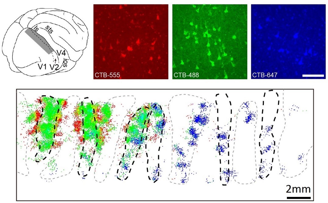
Fig. 1. Retrogradely labeled neurons in V2 CO stripes in a monkey. Top: illustration of a macaque brain and regions of V2 (shading). Three right panels are single wavelength fluorescence images of example labelled V2 neurons after V4 injections. Scale bar: 0.1 mm. Bottom: all labeled neurons overlaid on CO stripe outlines. Labeled neurons are represented by colored dots.
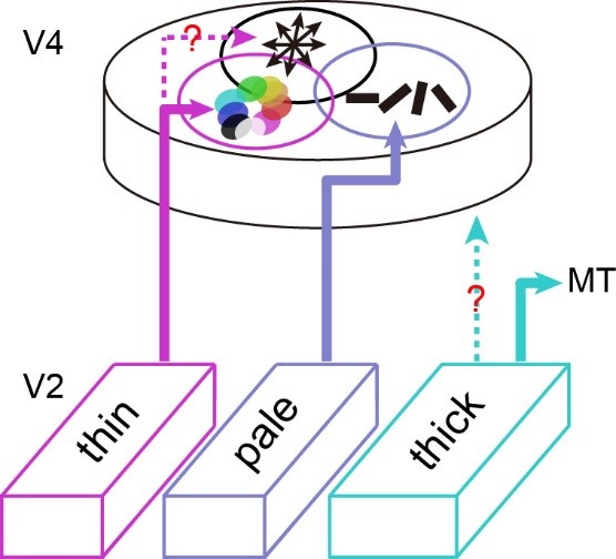
Fig. 2. Summary of functional inputs to V4 from V2. In V2–V4 projections, V4 color domains mainly receive inputs from V2 thin stripes, whereas V4 orientation domains mainly receive inputs from V2 pale stripes. Although we have not yet clearly determined whether V4 direction domains receive inputs from V2 thin stripes, they do not systematically receive inputs from thick or pale stripes. Some weak and unbiased projections from thick stripes to V4 may be due to uncertainty in identifying stripe types based on CO images. Neurons in thick stripes mainly project to area MT in the dorsal pathway.
Ph.D. candidates Chen Fang and Kun Yan from Dr. Haidong Lu’s lab are the co-first-authors of this paper. Lab members Chen Liang, Jiayu Wang, Xingya Cai and Rui Zhang contributed to this research. Professor Xiaohui Zhang, Yousheng Shu provided valuable technique supports. Other lab members also participated data collection. This work was funded by National Natural Science Foundation of China.
Link to the article:
Fang C*, Yan K*, Liang C, Wang J, Cai X, Zhang R, Lu HD (2022) Function-specific projections from V2 to V4 in macaques. Brain Structure and Function. (* equal contribution)
https://doi.org/10.1007/s00429-021-02440-3
Relevant studies from the lab:
Ma H, Li P, Hu J, Cai X, Song Q, Lu HD (2021) Processing of motion-boundary orientation in macaque V2. eLife. 10:e61317.
Tang R, Song Q, Li Y, Zhang R, Cai X, Lu HD (2020) Curvature-processing domains in primate V4. eLife. 9:e57502
Fang Y, Chen M, Xu H, Li P, Han C, Hu J, Zhu S, Ma H, Lu HD (2019) An orientation map for disparity-defined edges in area V4. Cerebral Cortex. 29:666-679.
Hu J, Ma H, Zhu S, Li P, Xu H, Fang Y, Chen M, Han C, Fang C, Cai X, Yan K, Lu HD. (2018) Visual Motion Processing in Macaque V2. Cell Reports. 25, 157–167.
Li P, Zhu S, Chen M, Han C, Xu H, Hu J, Fang Y, Lu HD (2013) A motion direction preference map in monkey V4. Neuron. 78:376–388.
Dr. Lu’s lab:
http://lulab.bnu.edu.cn/